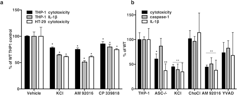
Figure 7. Inhibition of K+ channel activity blocked amebic cytotoxicity in HT-29 IECs and amebic cytotoxicity and IL-1β production in THP-1 macrophages.

(a) IL-1β secretion was measured by ELISA in HT-29 cells and differentiated THP-1 macrophages exposed to E. histolytica for 180 minutes (1 trophozoite to 5 cells). HT-29 cells did not secrete a detectable level of IL-1β (data not shown). Inhibitors did not cause LDH release in the absence of E. histolytica. Experimental values were normalized and expressed as the percent of vehicle treated controls, the mean and s.e.m. of three biological replicates is shown. P values were calculated relative to untreated cells (*P < 0.05; **P ≤ 0.008) by two-tailed Fisher’s LSD test. (b) LDH, IL-1β and cleaved caspase-1 in cells treated with KCl (50 mM), ChoCl (50 mM) AM92016 (10 μK) and YVAD (10 μ). Each experimental condition was normalized and is expressed relative to untreated wild type cells; the mean and s.e.m. of three biological replicates is shown. P values were calculated relative to untreated cells (*P ≤ 0.02; **P ≤ 0.005) by Fisher’s LSD test.
