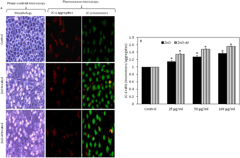
Figure 7. MMP loss due to pure and Al-doped ZnO nanoparticles exposure in MCF-7 cells.
(A) Representative fluorescent image showing that pure and Al-doped ZnO nanoparticles increased the JC-1 monomers (green fluorescence) and decreased the JC-1 aggregates (red fluorescence) in MCF-7 cells as compared to control. Fluorescent images were captured with a fluorescence microscope (OLYMPUS CKX 41). Morphology of cells also supported the data of apoptosis (MMP loss) showing that treated cells detached from surface and become rounded. (B) JC-1 ratio (monomers/aggregates) in treated and control cells. Data represented are mean ± SD of three identical experiments made in three replicate. *Statistically significant difference as compared to the controls (p < 0.05).

