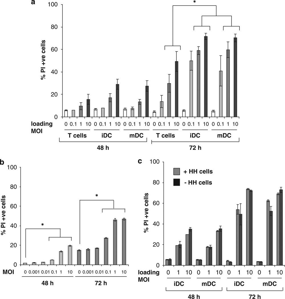
Figure 1.
T cells and DC are able to deliver reovirus for melanoma killing in vitro. (a) C57Bl/6 splenocytes, iDC and mDC were loaded with reovirus at the MOI indicated for 4 h at 4 °C, then washed two times in PBS and mixed with B16 melanoma targets at a 1:1 ratio. At 48 and 72 h the B16 cells were harvested and stained with PI for FACS analysis. Graphs show the mean±s.e. of data from three independent experiments; * denotes the significance of P<0.05. (b) Then, 105 B16 melanoma cells were cultured with reovirus at 0, 0.001, 0.01, 0.1, 1, or 10 p.f.u. per cell for 48 or 72 h, before being harvested. Cell death was determined by FACS analysis after staining the cells with PI. Graphs show mean±s.e. of data from five independent experiments, * denotes the significance of P<0.05. (c) iDC and mDC were loaded with reovirus at the MOI indicated for 4 h at 4 °C then washed two times in PBS and mixed with B16 targets at a 1:1 ratio. After incubation overnight, the hitchhiking cells were either removed or not, and fresh medium was added. After 48 and 72 h the B16 cells were harvested and analysed by PI staining. Graphs show mean±s.e. of data from two independent experiments.

