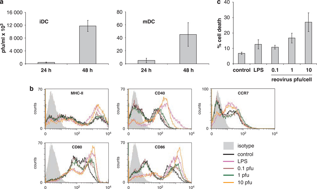
Figure 2.
The effects of reovirus on murine BMDC. (a) DC were loaded with reovirus at MOI 10 then washed, re-suspended in medium and cultured for 24 or 48 h before being harvested, re-suspended in PBS, freeze-thawed and the presence of reovirus estimated by standard plaque assay. Graphs show the mean±s.e. of data from two independent experiments. (b and c) Then, 3 × 10⁶ BMDC were cultured with 0, 0.1, 1, 10 p.f.u. per cell reovirus, or 100 ng ml−1 LPS. (b) MHC-II, CD40, CD80, CD86 and CCR7 expression after 24 h culture; data shown is representative of three independent experiments. (c) BMDC cell death after 48 h culture as determined by PI staining and FACS analysis. Graph shows mean±s.e. of data from five independent experiments.

