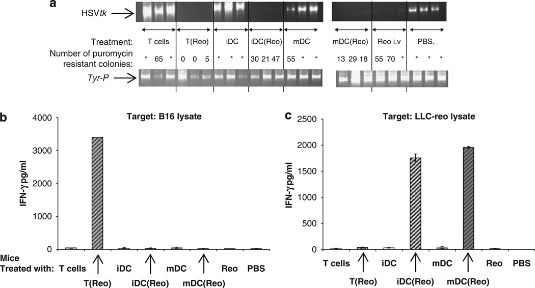
Figure 4.
T cells carrying reovirus prime anti-tumour immunity in naive mice. C57Bl/6 mice (three per group) were seeded s.c. with 5 × 10⁵ B16tk cells. After 10 days the mice were treated with PBS, 2 × 10⁶ p.f.u. reovirus or 2 × 10⁶ CTB-labeled Tcells, iDC or mDC loaded at MOI of 0 or 1. After a further 10 days the TDLN were explanted, dissociated and screened by PCR for the presence of the HSVtk and Tyr-P (control) genes (a); in addition, the dissociated TDLN were plated in puromycin-containing medium and the number of puromycin-resistant colonies were counted (=over 100 colonies)—results shown are for individual mice; P<0.05 for T(reo), iDC(reo) and mDC(reo) compared with controls. In addition, P<0.05 for T(reo) and mDC(reo) compared with neat virus, and for T(reo) compared with iDC(reo) and mDC(reo). (b and c) Pooled splenocytes from the same mice were cultured for 48 h with B16 lysate or reovirus-infected Lewis lung carcinoma (LLC-reo) lysate and the culture supernatants were assayed for the presence of IFN-γ by ELISA. Graphs show mean±s.e. of triplicate wells.

