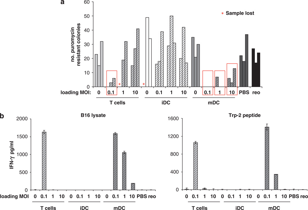
Figure 6.
T cells and mDC carrying reovirus prime anti-tumour immunity in virus-immune mice. Mice (three per group) were immunized with 108 p.f.u. reovirus i.p. After 2 weeks the mice were seeded s.c. with 2 × 10⁵ B16tk cells. After a further 10 days, the mice were treated with PBS, 2 × 10⁷ p.f.u. reovirus or 2 × 10⁶ T cells, iDC or mDC loaded at MOI of 0 or 0.1, 1 or 10. 10 days later the TDLN and spleens were explanted. Dissociated LN cells were cultured in medium containing puromycin and the number of puromycin-resistant colonies were counted (a); bars represent individual mice and show the number of puromycin-resistant colonies per 500 000 LN cells; red boxes denote significance compared with PBS control (P<0.05). (b) Pooled splenocytes were cultured for 48 h in the presence of the indicated peptide or cell lysates, and the culture supernatants were assayed for IFN-γ by ELISA; graphs show mean±s.e. of triplicate wells.

