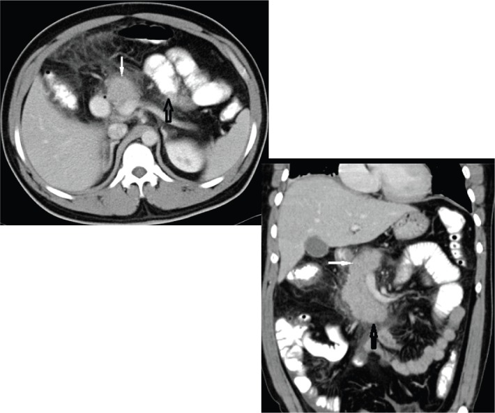
Figure 1.

Contrast-enhanced axial computed tomography (CT) [ Fig. 1(a)] and coronal CT [Fig. 1(b)] showing normal head (white solid arrow) and uncinate process (black solid arrow) with absence of distal neck, body and tail of the pancreas. Jejunal loops of small intestine (hollow black arrow) and stomach in the distal pancreatic bed can be seen (dependent stomach/dependent intestine sign).
