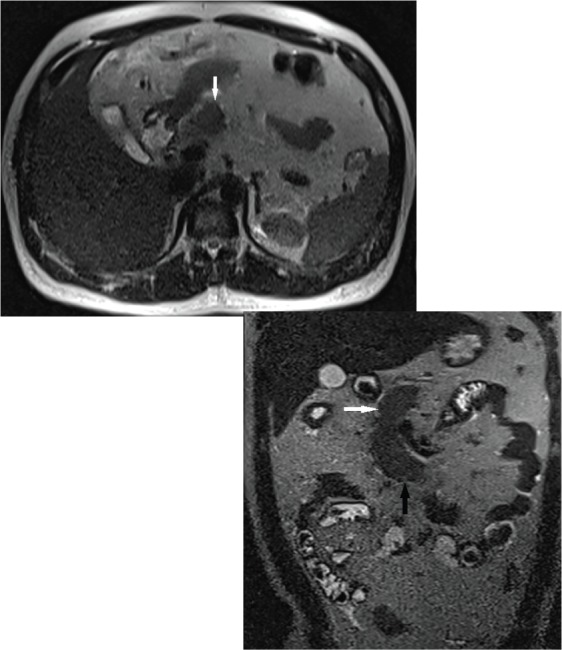
Figure 2.

Axial T2W magnetic resonance (MR) [ Fig. 2(a)] and coronal T2W MR [ Fig. 2(b)] images showing partial visualization of the pancreas. Well-developed, normal head (white solid arrow) and uncinate process (black solid arrow) of pancreas are visible, but the neck, body, tail and dorsal pancreatic duct cannot be seen.
