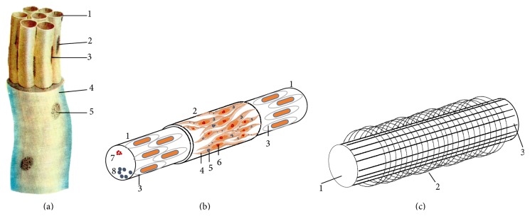
Figure 1.

Illustration of the primo-vessel and p-subvessel. (a) Primo-vessel. 1: primo-subvessel; 2: cell nucleus of the outer membrane; 3: nucleus of endothelial cell; 4: external jacket of primo-vessel; 5: nucleus of jacket endothelial cell [7]. (b) Diagram of primo-subvessel. 1: wall of subvessel formed by endothelial cells; 2: outer membrane of subvessel; 3: endothelial cell with rod-shaped nucleus; 4: spindle-shaped cell with ellipsoidal nucleus; 5: fine basophil granules in the cytoplasm; 6: fine chromatin granules inside nucleus; 7: basophil granules inside the subvessel; 8: p-microcells. (c) Diagram of subvessel fibers. 1: primo-subvessel; 2: fine transversal fiber; 3: longitudinal fiber.
