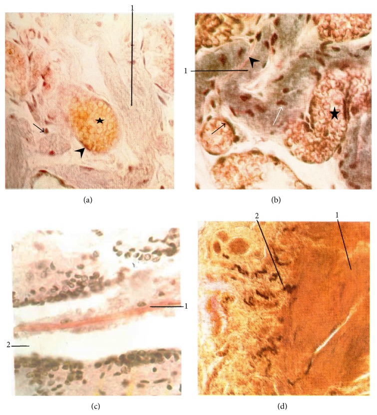
Figure 4.
Fibers. (a) Superficial primo-node (resorcin-fuchsin stain) (×400). 1: sinus, arrow: elastic fiber, arrowhead: elastic membrane in blood vessel, and star: erythrocytes. (b) Superficial primo-node (Verhoeff stain) (×400). 1: sinus, white arrow: basophil particle, star: erythrocytes, blood vessel membrane: black arrow, and collagen fiber between sinus folds: arrowhead. (c) Neural Bonghan duct (in the central canal of the spinal cord) (Van Gieson stain) (×400). 1: primo-vessel, 2: central canal of the spinal cord. (d) Nerve-supply at the superficial primo-node (Gros-Schultze reaction) (×160). 1: superficial primo-node, 2: nerve fiber [7].

