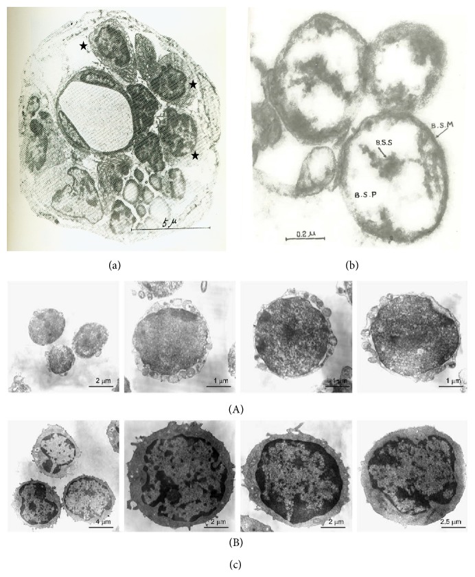
Figure 7.
TEM micrographs of p-microcells, very small embryonic-like (VSEL), and hematopoietic stem cells. (a) External primo-node. Star: p-microcells. (b) P-microcells. BSS: p-microcell nucleosome, BSP: p-microcell nucleoplasm, and BSM: p-microcell membrane [7]. (c) TEM of very small embryonic-like (VSEL) cells and hematopoietic stem cells. (A) Small embryonic-like (VSEL) cells are small and measure 2–4 μm in diameter. They possess a relatively large nucleus surrounded by a narrow rim of cytoplasm. The narrow rim of cytoplasm possesses a few mitochondria, scattered ribosomes, small profiles of endoplasmic reticulum, and a few vesicles. The nucleus is contained within a nuclear envelope with nuclear pores. Chromatin is loosely packed and consists of euchromatin. (B) In contrast hematopoietic stem cells display heterogeneous morphology and are larger. They measure on average 8–10 μm in diameter and possess scattered chromatin and prominent nucleoli (reprinted by permission from Macmillan Publishers Ltd. leukemia [36], copyright, 2006).

