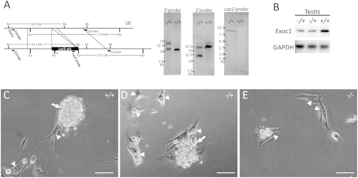
Figure 6. Exoc1 knockout mouse.
(A) Southern blotting of Exoc1−/+ and Exoc1+/+ mice. MfeI-digested DNA fragments were detected by 5′ outer and LacZ inner probes, and the BamHI-digested DNA fragments were detected by 3′ outer probes. (B) Western blotting showed that Exoc1 protein expression was reduced in Exoc1−/+ mice. (C–E) Cultivated blastocysts from Exoc1−/+ intercrosses. TG-like cells with an enlarged nucleus (arrowheads) were seen in cultivated embryos of either genotype. Dome-shaped colonies (arrows) were not seen in Exoc1−/−. M, MfeI; Ba, BamHI; Scale bar = 100 μm.

