
Figure 3. Surface representation of the hydrophobic residues located in the C-, D-, E-strands and CD- and DE-loop regions (residues 36 to 69) of the wild-type β2m (a), D76N (b), D59P (c), and W60C (d) in their representative IT-state conformations.
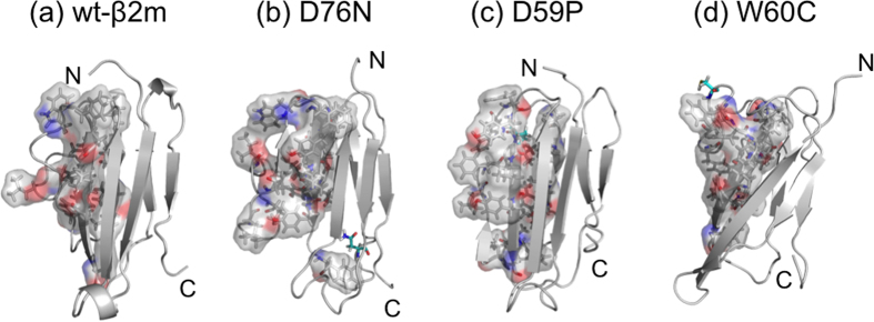
The mutation sites are indicated with cyan stick representation.