
Fig. 3.
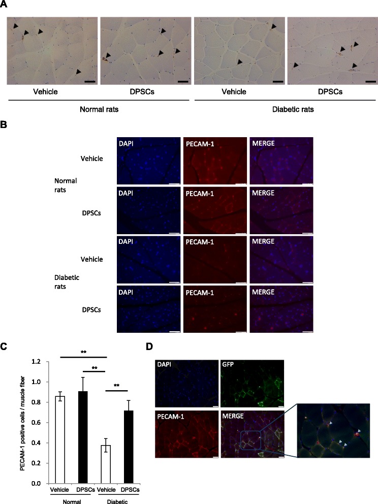
Capillaries in the soleus muscles. a, b Representative photomicrographs of the histological sections in the skeletal muscles of normal and diabetic rats. Capillaries were visualized by von Willebrand factor (vWF) (a) and PECAM-1 (b). Arrows indicate vascular endothelial cells stained by vWF. Bar = 50μm. c Quantitative analysis for capillary to muscle fiber ratio of the skeletal muscles of normal and diabetic rats. Results are expressed as means ± SEM (n = 4). **P < 0.01. d Differentiation of transplanted green fluorescent protein (GFP)-expressing dental pulp stem cells (DPSCs) into vascular endothelial cells in the skeletal muscles 4 weeks after the transplantation. DPSCs from GFP-expressing rats were transplanted into hindlimb skeletal muscles in the diabetic rats. Vascular endothelial cells were visualized by PECAM-1. Bar = 50 μm