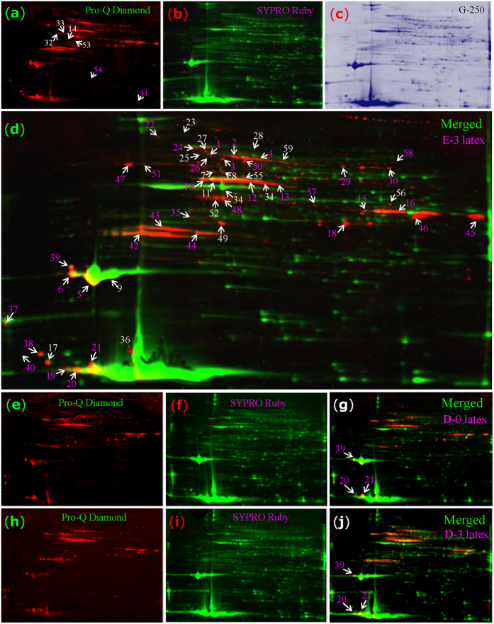
Figure 5. Phosphoproteomics analysis of rubber latex proteins upon ethylene treatment.
The 2-DE gel for E-3 latex was stained with Pro-Q Diamond dye to detect phosphoproteins (a, red). It was then restained with SYPRO Ruby (b, green). The same gel was restained by G250 (c, blue). The combination image of Pro-Q Diamond and SYPRO Ruby are presented to demonstrate the specific phosphorylated proteins (d, merged). The latex proteins obtained from the D-0 (e-g) and D-3 (h-j) plants were also stained by Pro-Q Diamond and SYPRO Ruby. Finally, 59 phosphoproteins (P1-P59) were identified by MS (d).

