
Fig. 1.
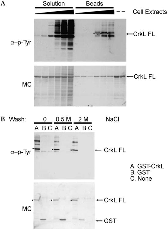
Bcr-Abl tyrosine kinase assays in solution and immobilized on beads. (A) GST-CrkL was incubated with 5, 10, 50, 100, 200, and 400 μg of K562 cell extracts in solution-phase and solid-phase kinase assays as described in the Materials and Methods. Specific quantities of the reaction mixture from solution reactions, the elution product from the solid-phase assays, and GST-CrkL alone were loaded onto a 12% SDS–PAGE gel. Following electrotransfer, the membrane was treated with Memcode reversible protein stain (MC, lower panel) to confirm equal loading and transfer, immunoblotted with 4G10 monoclonal antiphosphotyrosine antibody and horseradish peroxidase-linked secondary antibody, and then imaged with enhanced chemiluminescence (α-p-Tyr, upper panel). FL, full-length. (B) Comparison of Bcr-Abl tyrosine kinase assays with GST-CrkL or GST bound to glutathione beads or the beads alone suggests that background immunoreactivity results from phosphoproteins bound to the CrkL moiety. Following incubation with cell extract and washing as before, beads were washed with 0, 0.5, or 2 M NaCl in 50 mM Tris-HCl (pH 7.5), respectively, before elution and Western analysis. Lower panel: Memcode protein stain (MC). Upper panel: 4G10 antiphosphotyrosine antibody (α-p-Tyr). FL, full-length.