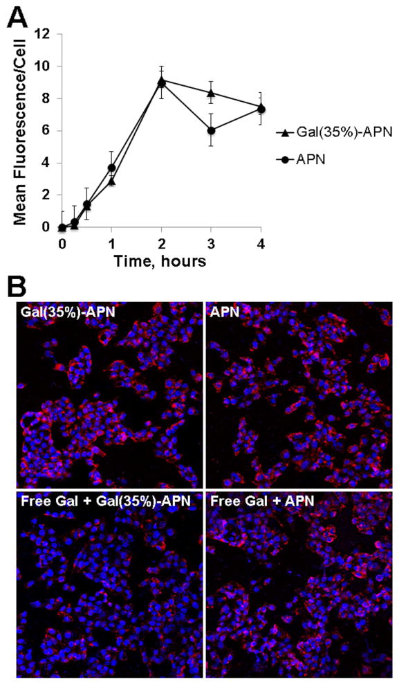
Figure 2.
Uptake of Gal-APN into HepG2 cells. (A). Time-dependent uptake of Gal(35%)-APN and APN at 37°C as quantified from confocal images (objective 20x). Data are presented as mean ± SEM (n = 15–20). (B). Representative confocal images of HepG2 cells incubated with Cy5-labeled Gal(35%)-APN and APN in the absence or presence of free galactose. For competition experiments, cells were pre-treated for 0.5 h with 100 mM of galactose followed by co-incubation with corresponding APN for 1 h. The merged images of Cy5-labeled p41 (red) and cell nuclei stained with DAPI (blue) (objective 20x). In all experiments, cells were treated with APN at a dose equivalent to 5 μM peptide at 37°C.

