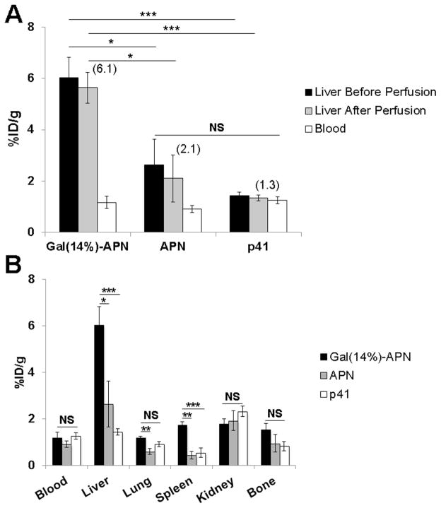
Figure 5.
Biodistribution of radiolabeled Gal-APN in vivo at 4 h post i.v. injection. (A) Quantification of the liver (before and after perfusion) and blood concentrations of the Gal(14%)-APN, APN and p41 (50 μg p41 or its APN format per animal). The values in parenthesis indicate the mean liver (perfused)-to-blood ratio. (B). Tissue distribution of Gal(14%)-APN, APN and p41. Apart from liver, a higher accumulation of Gal(14%)-APN was found in lung and kidney. All results were interpreted as mean percentage of injected dose per gram of tissue (%ID/g) ± SEM (n = 6); *p < 0.05, **p < 0.01, ***p < 0.001, NS - not significant.

