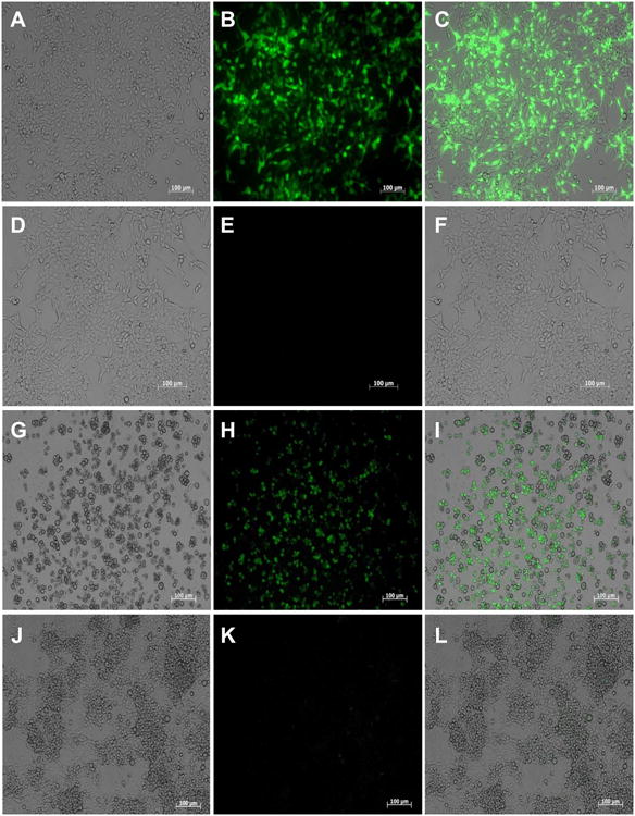
Fig. 5.

GFP expression in cells transfected with the plasmid piLenti-RNAi-GFP and polyethylenimine (PEI). (A – F) Representative images of transfected human cells (HEK 293T) examined under bright field (A), fluorescent field (B) and merged (C). PEI only-transfected control human cells examined either under bright (D), or fluorescence field (E) and merged (F). Pictures of HEK 293T cells were captured 18 h after transfection, (G – L) Representative images of transfected Bge (Biomphalaria glabrata embryonic) cells examined under either bright (G), or fluorescence field (H) and merged (I). PEI only-transfected Bge cells examined under either bright (J), or fluorescence field (K) and merged (L). Pictures of Bge cells were captured 5 days after transfection. Scale bar: 100 μm.
