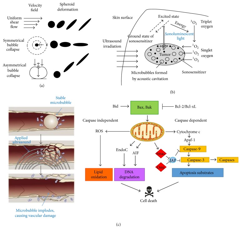
Figure 1.
Antineoplastic mechanisms of ultrasound. (a) Microbubbles are unevenly stretched by ultrasonic waves, causing an unequal distribution of force known as inertial cavitation. Microbubbles oscillating in a stable motion reflect stable cavitation, while the expansion and contraction of microbubbles that are unequal and markedly exaggerated are indicative of inertial cavitation. Subsequent stress results in microbubble implosion, creating considerable amounts of energy. (b) The energy provided by the collapse of microbubbles potentiates the formation of sonoluminescent light within the cell. The light subsequently activates endogenous compounds within the cell that release ROS when returning to the ground state. (c) Many tumors rely on angiogenesis to sustain increased metabolic activity. Microbubbles can enter the tumor vasculature, and at sufficiently high amplitudes, ultrasound induces significant vascular damage, shutting down blood flow. The vessels develop and harbor hypoxic regions, causing oxidative stress; lack of nutrients and increased acidity induce apoptosis. In addition, malignant cells exposed to ultrasound often undergo apoptosis through the intrinsic pathway. Caspase-3 is upregulated by proteins such as Bax and Bak that integrate into the mitochondrial membrane, facilitating apoptotic signaling. It is important to note that sonosensitizers have been developed to significantly increase the efficacy of each mechanism. Images courtesy of [1].

