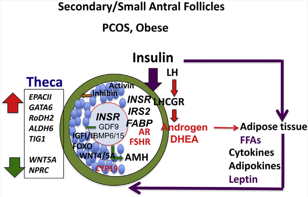
FIGURE 3.
Transition from primary to secondary/small antral follicles. In obese PCOS patients, hyperinsulinemia from adiposity-dependent insulin resistance and altered adipogenesis impair follicular development by mechanisms that appear to differ from those in lean PCOS patients. Insulin may have a greater influence than LH on theca, granulosa, and likely oocyte functions. Adipokines also likely alter each cell type within the follicle. Note that insulin receptor (INSR) expression may be elevated in oocytes of PCOS patients compared to non-PCOS patients (205). Cumulus cells of obese PCOS patients also express higher levels of INSR and specific fatty acid binding proteins (FABPs) (102). Theca cells produce elevated levels of androgens, especially DHEA, in response to LH and elevated expression of CYP17A1 and CYP11A1, which are targets of GATA6 and retinoic acid (RA) (47). The PCOS theca cells exhibit increased expression of enzymes controlling RA production and enhanced responses to RA, including steroidogenesis and the target gene TIG1. These responses may be related to cAMP signaling through exchange protein activated by cAMP II (EPAC II). Conversely, PCOS theca cells exhibit reduced expression of WNT signaling and cGMP signaling pathways, but the functional significance of this remains to be determined. Oocyte factor and GDF9 signaling is denoted by green arrows; inhibin by the orange arrow; LH and androgen signaling by red arrows and letters; and insulin signaling by purple arrows and letters.

