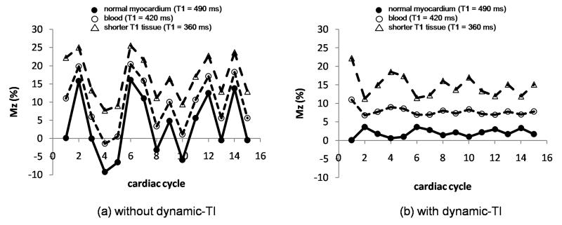
Figure 3.
Longitudinal magnetization, Mz (as a percentage of the initial longitudinal magnetization, M0) of blood (open circles, T1 = 420 ms) and myocardium (filled circles, T1 = 490 ms) for the most central k-space line in an inversion prepared segmented gradient echo acquisition with single R-wave gating without (left) and with (right) dynamic-TI in a subject with variable heart rate. A shorter T1 tissue is also simulated (open triangles, T1 = 360 ms). The sequence repeat time intervals were from an example patient (Figure 4): 633ms, 966ms, 1433ms, 1266ms, 633ms, 699ms, 1083ms, 850ms, 1216ms, 833ms, 683ms, 983ms, 666ms, 983ms. The first two cardiac cycles were dummy cycles. The longitudinal magnetization at the centre of k-space is less variable for all T1s with the dynamic-TI algorithm.

