
Figure 1.
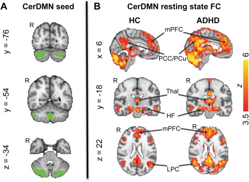
CerDMN seed and its FC in the HC and ADHD groups. (A) The CerDMN seed (based on [Buckner et al., 2011]) from which the resting state fMRI time series were extracted. B) Voxels showing significant positive FC with the CerDMN in the HC and ADHD groups [FWE‐corrected Z > 2.3 (threshold increased to 3.5 for display purposes); cluster‐based P < 0.05]. HF, hippocampal formation; LPC, lateral parietal cortex; mPFC, medial prefrontal cortex; PCC, posterior cingulate cortex; PCu, precuneus; Thal, thalamus. [Color figure can be viewed in the online issue, which is available at http://wileyonlinelibrary.com.]