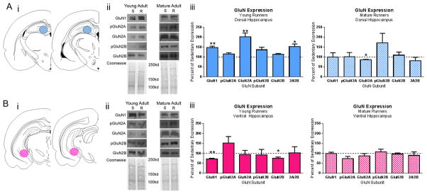
Figure 3. GluN Expression in the Dorsal and Ventral Hippocampus of Young Adult and Mature Adult Runners.

Dorsal (A) and ventral (B) hippocampus tissue was collected from young and mature adult runners. i) Schematic representations of dorsal (AP −3.14 mm to −4.30 mm from bregma) and ventral hippocampus (AP −5.3 mm to −6.1 mm from bregma) sections adapted from (Paxinos and Watson, 2007). Tissue punches were collected from 500 μm thick sections of young adult and mature adult sedentary and running rats. Blue circles represent site of dorsal tissue collection and pink circles represent site of ventral tissue collection via tissue punch. ii) Representative western blots and associated coomassie staining in sedentary (S) and running (R) animals. iii) Summarized data for GluN subunit expression as percent of sedentary age-match controls. Asterisks denote significant differences between age groups of runners within hippocampal subregion. *p≤0.05, **p≤0.01
