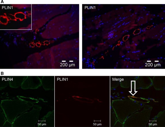
Figure 6.

Intramuscular adipocytes. (A) Sections of paraffin-embedded biopsies from m. vastus lateralis (n = 8) were stained with PLIN1 antibody (red, mouse anti-PLIN1) to detect adipocytes (A). Representative images (200X) were obtained by wide-field fluorescence microscopy. High magnification (1000X) of adipocytes is shown in the insert. (B) The sections were stained for PLIN4 (green, rabbit anti-PLIN4) and PLIN1 (red, mouse anti-PLIN1), showing partly co-localization in intramuscular adipocytes (n = 4). The merged image (200X) shows co-localization of the proteins (white arrow). The image was obtained by confocal fluorescence microscopy.
