
Figure 7.
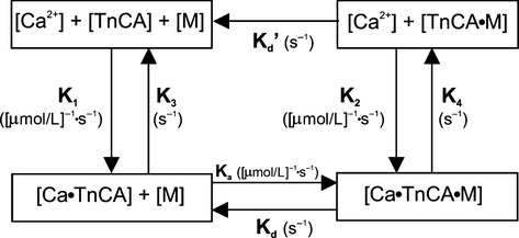
Block diagram of a biochemical model relating the input/output relationship between myoplasmic [Ca2+] and LVP adapted from Baran et al. (1997) and Shimizu et al. (2002) The four-state model is governed by five differential equations. TnCA represents the troponin C molecule on the actin (A) myofilament, M represents the myosin head, + indicates weak bonds and • represents strong bonds. The sequence of events from phasic [Ca2+] to contraction are as follows: Ca2+ binds to TnCA, tropomyosin shifts so M and A can bind forming an actinomyosin cross-bridge, Ca2+ dissociates from TnCA with cross-bridge attached, and finally the cross-bridge breaks. Note that A and M cannot form cross-bridges in the absence of Ca2+; however, since this is a loose coupling model, once a cross-bridge has been formed it no longer requires associated Ca2+ to remain attached. Adapted from Rhodes et al. (2006a,b). Model rate constants and their units are indicated by their sites of action and described in Table1. Model parameter values for stretch before and after stunning are given in Table2.