
Figure 5.
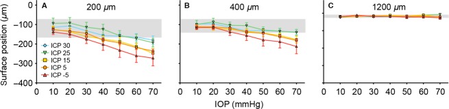
Effect of ICP on IOP-induced changes in surface deformation. Panels (A–C) (n = 6–7) show raw surface deformation at (A) the optic nerve head 200 μm from midline. (B) the central retinal 400 μm from midline and (C) the peripheral retina 1200 μm from the midline. (mean ± SEM). Blue diamond: 30 mmHg; green down triangle: 25 mmHg; yellow square: 15 mmHg; orange circle: 5 mmHg; red up triangle: −5 mmHg. Gray area: 95% confidence interval of the baseline (IOP = 10 mmHg).