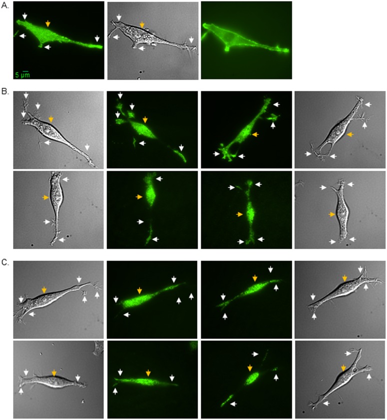
Fig 2. Spatial arrangement of glycosylated and unglycosylated forms of Kv proteins in B35 cells.
Microscopy images were acquired in TIRF (left panel), DIC (middle panel), and wide-field (right panel) modes of EGFP tagged wild type Kv3.1b heterologously expressed in B35 cells (a). TIRF images (middle panels), along with their accompanied DIC images (outer panels), are shown for wild type Kv3.1a (b, upper row) and Kv1.1 (c, upper row) proteins, and also for N220Q/N229Q Kv3.1a (b, lower row) and N207Q Kv1.1 (c, lower row) proteins. Representative scale bar (5 μm) was identical for all images. White and gold arrows point to EGFP tagged Kv proteins in the outgrowth and cell body, respectively.

