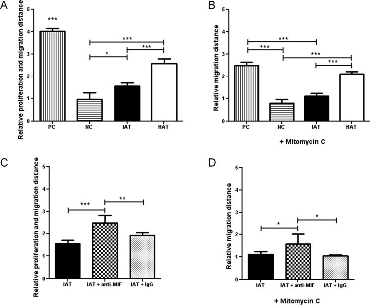
Fig 7. In vitro scratch assay of dermal fibroblasts and supernatants from IAT and HAT.
Wound healing of human dermal fibroblast monolayers was evaluated by an in vitro scratch assay. Fibroblasts were incubated with supernatants of samples from IAT and HAT, MIF-dependency was evaluated by anti-MIF antibodies. Medium containing 10% FCS served as a PC and medium containing 0% FCS served as a NC. Experiments were performed in the presence (A, B) and absence (C, D) of the proliferation inhibitor Mitomycin C. A Supernatants from IAT show a decreased induction of fibroblast proliferation and migration (no Mitomycin C). B Fibroblast proliferation and migration is increased by addition of MIF-antibodies (no Mitomycin C). C Supernatants from IAT show a decreased induction of fibroblast migration (10 μg/ml Mitomycin C). D Fibroblast migration is increased by addition of MIF-antibodies (10 μg/ml Mitomycin C). All measurements were normalized to the negative control of Fig 7A which was set as 1. Data are presented in relative migration distance of fibroblasts ± SEM. Statistically significant differences are indicated by asterisks (*, p<0.05; **, p<0.01; ***, p<0.001).

