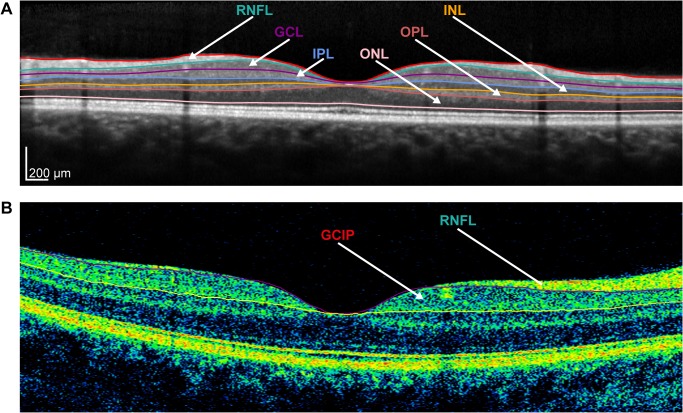
Fig 2. Intra-retinal layer segmentation of OCT B-scans.
(A) Exemplary B-scan of a Spectralis SD-OCT with manually reviewed and corrected segmentation into the following retinal layers: retinal nerve fiber layer (RNFL), ganglion cell layer (GCL), inner plexiform layer (IPL), inner nuclear layer (INL), outer plexiform layer (OPL) and outer nuclear layer (ONL). (B) Exemplary Cirrus HD-OCT B-scan with segmentation in the RNFL and combined ganglion cell and inner plexiform (GCIP) layer.

