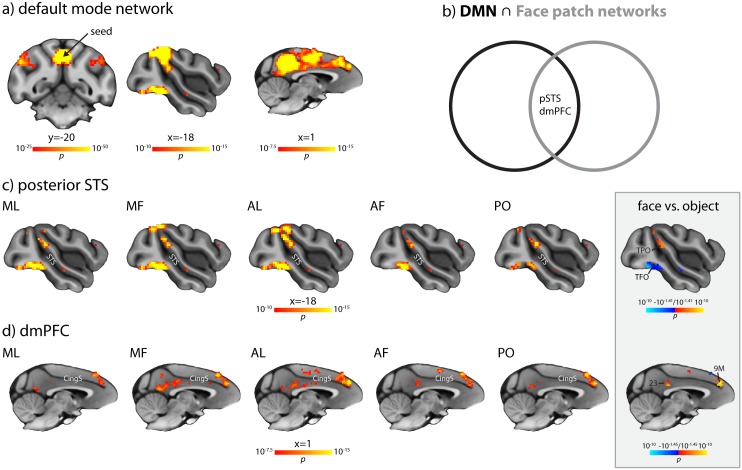
Fig 5. Overlap analysis of the DMN and FPRSNs.
(a) The classical monkey DMN on coronal and sagittal slices. The arrow indicates the approximate location of the DMN seed. Note that the sagittal slices are the same as in c and d, respectively, at corresponding statistical thresholds. (b) Illustration of the overlap analysis. In humans, the intersection between the DMN and the social brain isolates areas involved in high-level social cognition, such as TPJ in the posterior STS, dmPFC, and medial PPC [22–24]. We tested, using the FPRSNs as a proxy for the social brain, whether and where a similar overlap exists in the macaque. (c) Voxels in area TPO in the dorsal bank of the posterior STS, located dorsally of FST, laterally to MST, and anterior and dorsal of MT, that show significant connectivity both with the PPC and with the respective face patch (AF, AL, MF, ML, and PO) at p < 10−10, uncorrected. The corresponding Jaccard Indices are 0.1358 for AF, 0.1635 for AL, 0.1786 for MF, 0.1366 for ML, and 0.1150 for PO. Also evident is overlap around the occipitotemporal sulcus, which is not part of the classical DMN. The grey inset shows that the strength of connectivity to voxels in which AL showed overlap with the DMN in the posterior STS was significantly higher for AL than for the object patch. In contrast, connectivity to the occipitotemporal sulcus was significantly higher for the object patch than for AL. (d) Voxels in dmPFC (areas 9M/10) and medial PPC (areas PGm/23) that show significant connectivity both with the PPC and with the respective face patch (AF, AL, MF, ML, and PO) at p < 10−7.5, uncorrected. The corresponding Jaccard Indices are 0.2006 for AF, 0.2268 for AL, 0.2530 for MF, 0.2062 for ML, and 0.1821 for PO. The grey inset shows that the strength of connectivity to voxels in which AL showed overlap with the DMN in the dmPFC (area 9M) and medial PPC (area 23) was significantly higher for AL than for the object patch. Both inset maps are corrected for multiple comparisons with a FDR at q = 0.05, accounting for the number of voxels that show significant overlap between AL and the DMN at the same statistical thresholds as shown in panels c and d, respectively. All results are overlaid on the MNI-Paxinos template brain, in radiological convention (left is right). Coordinates are relative to the center of the anterior commissure. Area labels are based on Paxinos et al. [38]. See S10 Fig for overlap between the DMN and AM connectivity, which replicates the main findings at a more lenient statistical threshold.

