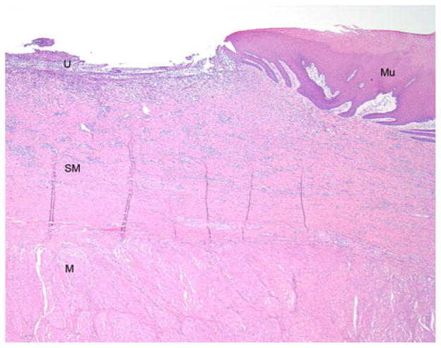
Fig. 10.
Photomicrograph (H&E, original magnification = 20×) from the edge of the stricture zone showing an ulcer (U) where the EEM occurred. The underlying fibroinflammatory reaction involves almost the entirety of the submucosa (SM), and the uninvolved muscularis propria (M) is visible beneath. At the edge of the ulcer, there is reepithelialization over the scar by reactive squamous mucosa (Mu)

