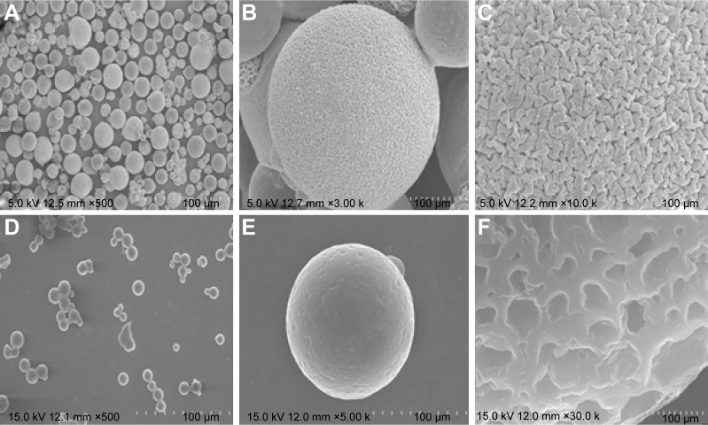
Figure 3.
SEM images of Fbg microspheres.
Notes: (A–C) Bare-Fbg microspheres, (D–F) DOX–linker 1–Fbg microspheres, (A and D) distributions of the microspheres, (B and E) morphology of the microspheres, (C and F) surface fine structure of the microspheres.
Abbreviations: SEM, scanning electron microscope; DOX, doxorubicin; Fbg, fibrinogen.

