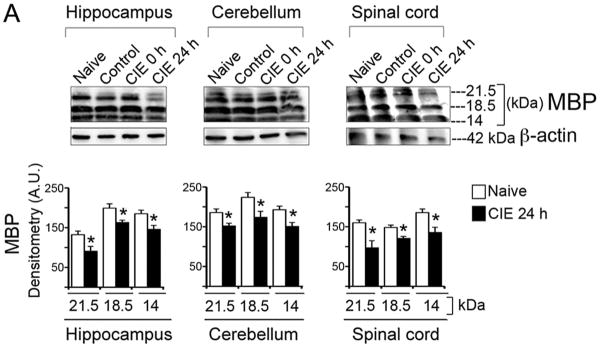
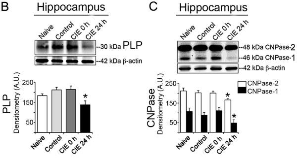
Fig. 2. CIE-induced loss of myelin proteins and enzyme in brain and spinal cord.


(A) Immunoblots and corresponding densitometric analysis (A.U., mean ± S.E.M.) of MBP showed significantly (*p < 0.001) reduced MBP isoforms (21.5, 18.5 and 14 kDa) in hippocampus, cerebellum and spinal cord in CIE 24 h (withdrawal) compared with the respective MBP isoforms in naïve mice (n = 4-6). Likewise significant reduction was seen in (B) PLP (30 kDa) and (C) CNPase-1 and-2 (46 and 48 kDa) in hippocampus in CIE 24 h (withdrawal) compared with naïve group of mice (*p < 0.0001; n = 4-6). Each immunoblot was re-probed for β-actin (42 kDa), which showed tendency towards marginal loss in CIE 24 h group compared to rest of the groups.
