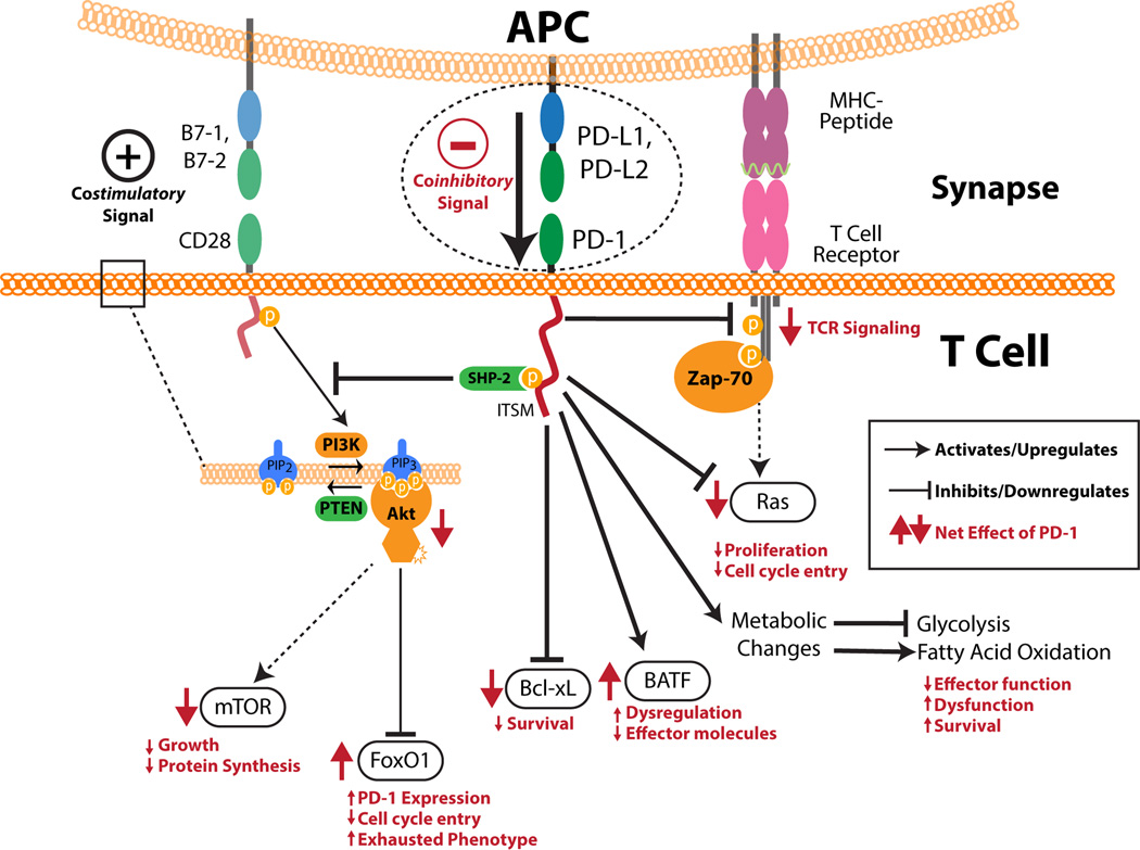
Figure 1. PD-1 signaling.
PD-1 has both an intracellular immunoreceptor tyrosine-based switch motif (ITSM) and immunoreceptor tyrosine-based inhibitory motif (ITIM) in its cytoplasmic tail. SHP-2 can bind to the phosphorylated ITSM. PD-1 ligation by ligands leads to overall inhibition of TCR signaling through inhibition of CD3ζ chain phosphorylation and Zap-70 association. PD-1 signaling causes the downregulation of both Ras and Bcl-xL which affect proliferation and cell survival, respectively. An increase in BATF can be seen which impairs the effector function of T cells. PD-1 also inhibits the phosphatidylinositol 3-kinase (PI3K)/Akt pathway by inhibiting the activation of PI3K. This has downstream effects including the downregulation of mechanistic target of rapamycin (mTOR) and an increased half-life of FoxO1. PD-1 signaling also influences the cell’s metabolism by inhibiting glycolysis and promoting fatty acid oxidation. Together, all of these effects cause T cells to become less proliferative, lose their effector functions, and take on an exhausted and dysfunctional phenotype. The net effect of PD-1 ligation on all of these processes is shown in red with arrow direction indicating upregulation and downregulation.

