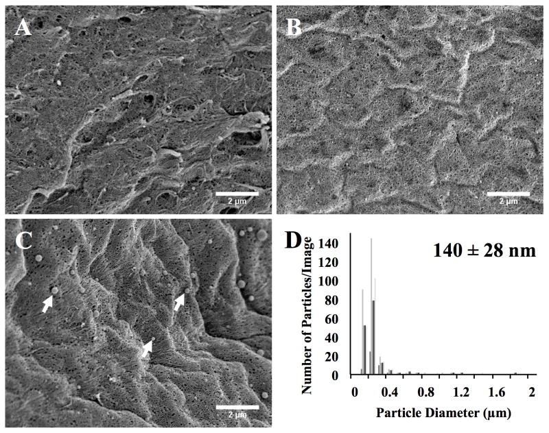
Figure 2.
FE-SEM images of the fibrin hydrogel surface after being treated for 4 hours with: (A) RNase-free water, (B) uncomplexed siRNA, or (C) Lipofectamine siRNA-GFP complexes (arrows). (D) ImageJ particle size analysis of adsorbed Lipofectamine siRNA-GFP complexes on the fibrin surface correlates with DLS analysis of solution suspension of Lipofectamine siRNA nanoparticles (numeric insert). ImageJ measurement of the diameter size distribution of the adsorbed siRNA complexes showed complex diameters centered near 200nm (15k x magnification; scale bar denotes 2μm).

