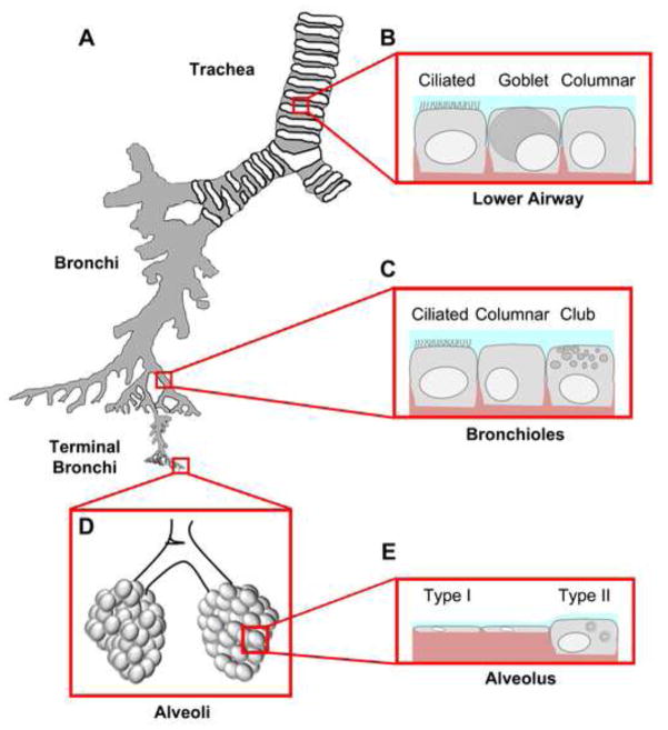
Figure 1. Epithelial diversity along the respiratory tree.
A. Airways are divided into four main segments: the trachea, the branching bronchi, the terminal bronchi, and the alveolar space. Each segment contains a unique mix of cell types that have specialized functions. B. The lower airway, proximal to the bifurcation of the left and right bronchus, consists of mostly ciliated cells whose main function is to sweep mucus secreted by goblet cells (blue) out of the airways. Columnar and other cells (e.g. serous cells) also contribute to the airway barrier. Basal cells (not shown) are localized to the basement membrane but do not contribute to the tight junction barrier. C. Distal to the tracheal bifurcation are bronchiolar cells that consist mainly of ciliated, columnar and club cells. Club cells secrete a specialized form of pulmonary surfactant as opposed to mucus and provide a transition zone between the airway and alveolar space. D. The alveolar space is the location of gas exchange and consists mainly of squamous type I and cuboidal type II cells. Tight junctions between these cells form at apical cell-cell interaction sites. The alveolar sac maintains surface tension through surfactant secreted by type II cells preventing alveolar collapse. E. Gas exchange occurs efficiently through type I cells, which make up the vast majority of alveolar surface area.

