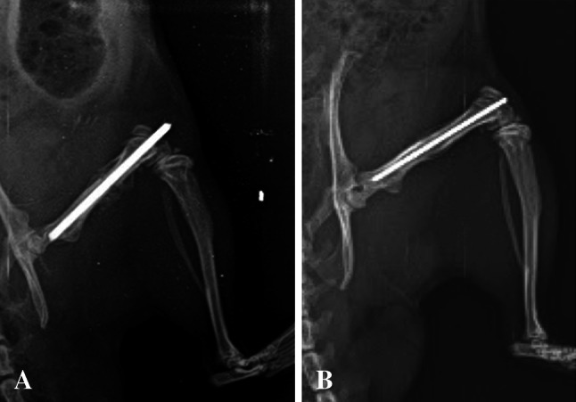
Fig. 1A–B.

Radiologic images at 6 weeks postoperatively are shown. Gross evaluation of radiographic image in the infection group reveals immature callus surrounding the fracture site and lack of bridging bone (A). Interval cortical bridging is seen in the control group (B).
