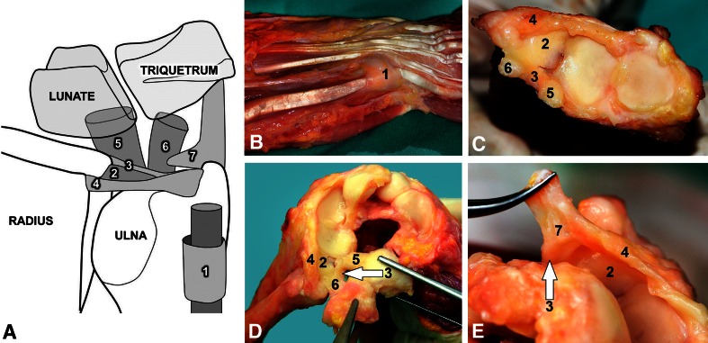
Fig. 1A–E.
(A) The triangular fibrocartilage complex consists of seven separate structures, namely the subsheath of the extensor carpi ulnaris tendon sheath (1 = SS-ECU), the articular disc (2 = AD), the volar radioulnar ligament (3 = VRUL), the dorsal radioulnar ligament (4 = DRUL), the ulnolunate ligament (5 = UL), the ulnotriquetral ligament (6 = UTq), and the ulnocarpal meniscoid (7 = UCM). (B) The subsheath of the extensor ulnaris tendon sheath is seen after removal of the extensor retinaculum. (C) Parts 2 through 6 of the triangular fibrocartilage complex are seen after removal of the carpal bones from the sagittal plane. (D) The ulnotriquetral ligament (6) inserts at the volar aspect of the triquetrum and originates on the radiovolar base of the ulna styloid. The ulnolunate ligament (5) inserts at the volar aspect of the lunate and originates mainly on the volar border of the articular disc (2). The dorsal radioulnar (4) and volar radioulnar (arrow 3) ligaments begin at the dorsoulnar or ulnovolar margin of the radius at the sigmoid notch and run toward the ulnar styloid. (E) A close-up view shows the ulnocarpal meniscoid (7).

