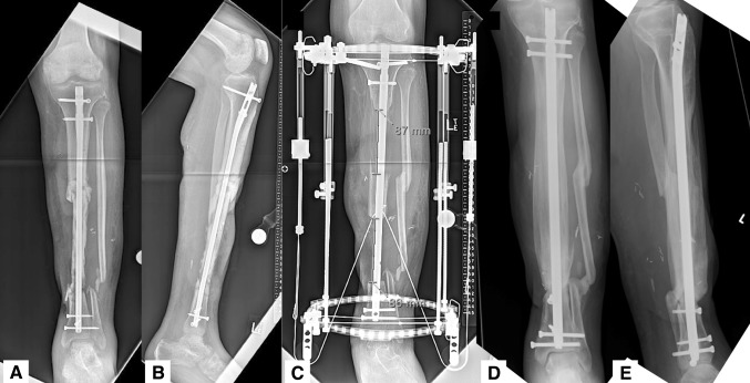
Fig. 1A–E.
(A) AP and (B) lateral radiographs show a 31-year-old man who presented with a 17.5-cm segment of infected tibial bone after a Type IIIB open tibia fracture. The patient was treated with soft tissue coverage, excision of nonviable bone, and bone transport over a nail (C). Final radiographs (D–E) demonstrate maintenance of alignment, docking site union, and no infection 1 year after reconstruction.

