
Figure 3.
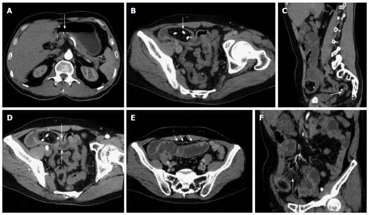
Representative case of a 58-year-old male with hawthorn small bowel obstruction. A: Contrast-enhanced examination result displaying the gastroduodenal anastomotic stoma of the artery; B: Oblique MPR revealing an oval bezoar in the distal end of the jejunum with shadows of high-density seeds (arrow); C: Sagittal reconstruction showing co-existing bezoar inside the ileum (arrow); D: Arterial phase image exhibiting the thickened and strengthened intestinal wall in the distal end of the obstruction site (arrow); E: Portal venous phase image showing a significantly enlarged vascular shadow and blurred mesentery of the proximal end of the dilated SBO intestine (arrow); F: CTA image revealing the thickening of the mesenteric blood vessel and peripheral exudation in the obstruction site (arrow).