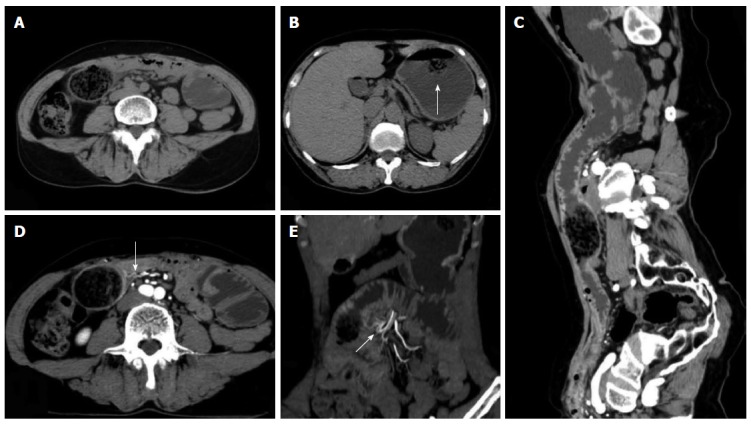
Figure 4.

Representative case of a 60-year-old female with diospyrobezoar-induced small bowel obstruction previously subjected to gastrectomy and gastrojejunostomy. A: Computed tomography (CT) planar scan image displaying the bezoar without an envelope inside the proximal end of the ileum; B: co-existing gastric bezoar with an envelope (arrow); C: CPR image showing the dilation of the proximal end of the obstructed intestine, bezoar and distal wall thickening at the obstruction site; D: Contrast-enhanced CT scan with an arterial phase image revealing a significant enhancement and thickening of obstruction regions and the distal end of the obstructed intestine; blood-supplying arteries exhibited hyperaemia (arrow); E: CTA image showing the same mesenteric artery (arrow) that provides blood supply for the transitional zone and the distal end of the obstruction site.
