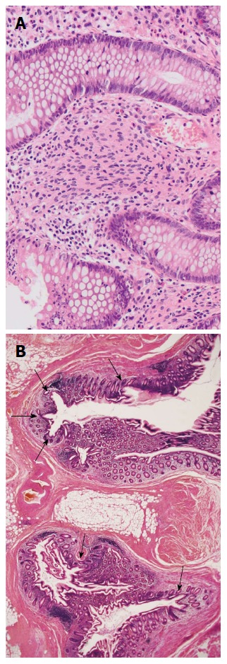
Figure 3.

Pathological findings (Hematoxylin-eosin staining). A: Spindle shaped tumor cells proliferated diffusely in the interstitium between the crypts, without forming a distinct mass (magnification × 10); B: Multiple diverticulums were present in the wall of the appendix. The majority of diverticulums were small in size, causing subtle epithelial herniation (magnification × 1).
