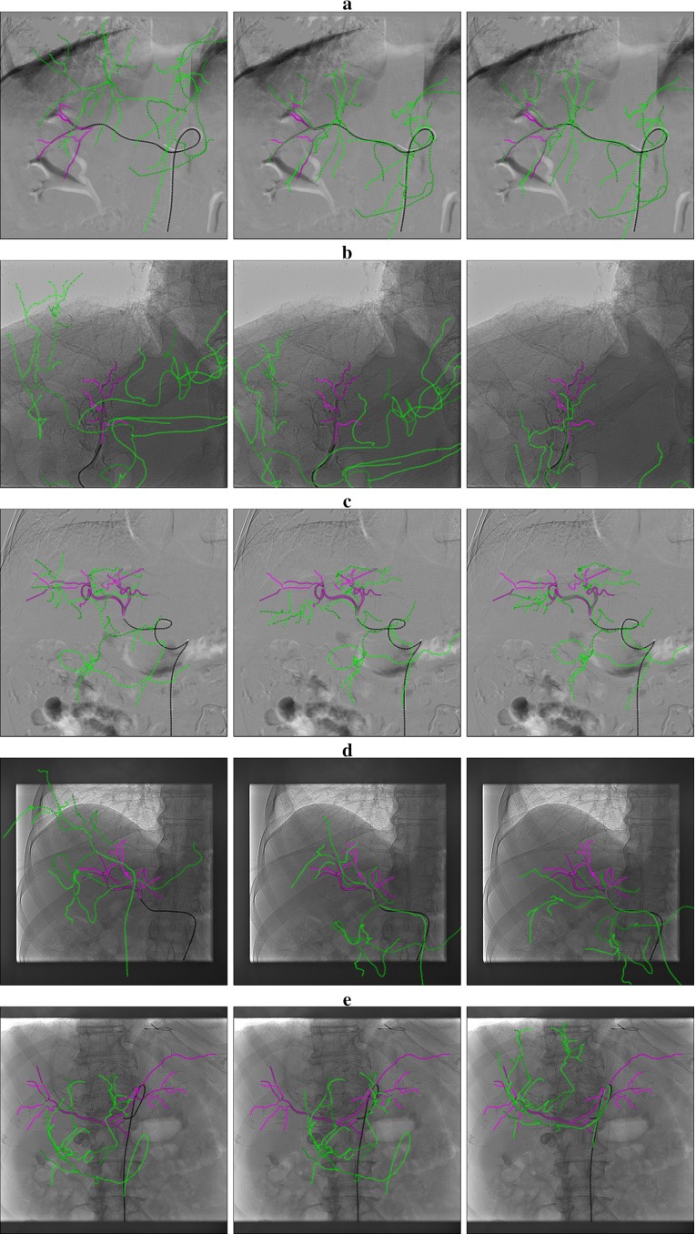
Fig. 16.
Projection of the 3DRA blood vessel (in green) with the catheter (in black) and the contrast agent (in purple). Initial position (left). Registered position with Powell (middle). Registered position with brute force (right). a The registration is correct. Here the catheter is long enough to give information. b The catheter part is too short. Powell registered with a good distance metric but the result is wrong. Brute force is correct. c The catheter tip position is correct for both optimizers. The vessels and catheter deformation prevent to have a perfect match. d Here the distance metric and the tip is correct with both optimizers but brute force rotates too much. e As a long part of the aorta is missing in the 3DRA, Powell stops in a local minimum while brute force is more exhaustive and reach the global minimum

