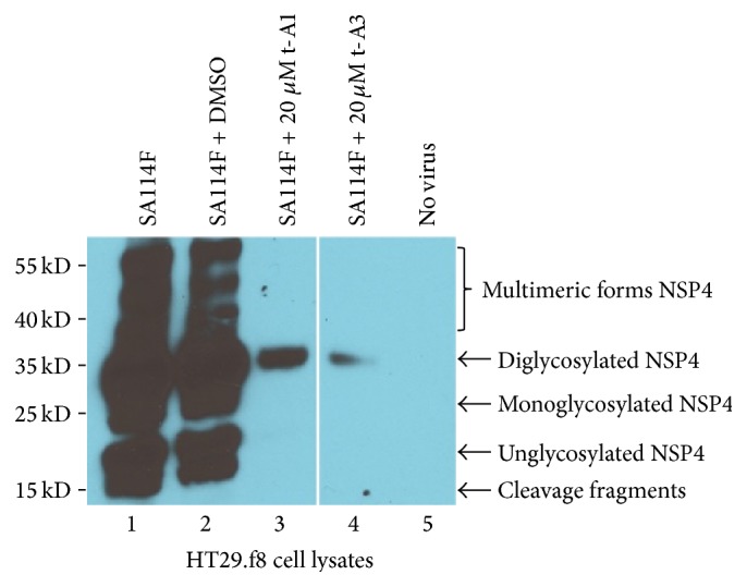
Figure 6.

Western blot analysis of HT29.f8 cell lysates. Five micrograms of HT29.f8 cell lysates was separated on a 12.5% SDS-PAGE, electroblotted onto nitrocellulose membranes, probed with rabbit anti-NSP4150–175 peptide-specific and goat anti-rabbit HRP-conjugated IgG, and visualized with Super Signal West Pico chemiluminescent substrate (Pierce) followed by exposure to Kodak X-OMAT film. (Lane 1) RV-infected HT29.f8 cells and (Lane 2) RV-infected HT29.f8 cells with 0.02% DMSO, respectively, show cleavage fragments and unglycosylated, mono-, diglycosylated, and multimeric forms of NSP4. (Lane 3) RV-infected HT29.f8 cells with 20 μM t-A1 and (Lane 4) RV-infected HT29.f8 cells with 20 μM t-A3 only show the diglycosylated form of NSP4. (Lane 5) HT29.f8 with no virus showed NSP4 banding pattern.
