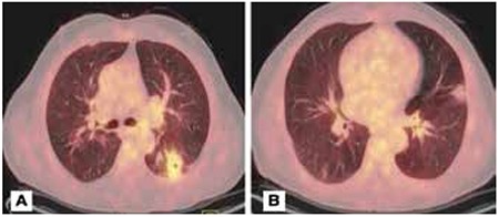
Figure 2. PET/CT scans of a patient with multiple nodules and cavitary lesionA) A cavitary lesion 2.5 cm in diameter in the superior segment of left lower lobe (SUVmax: 7.04). There is low metabolic activity in the left hilar lymph node (SUVmax: 3.38). B) Bilateral multiple nodules, the largest 2.5 cm in diameter (SUVmax: 3.93). The patient underwent trans-thoracic fine needle biopsy, which was reported as organizing pneumonia after pathologic evaluation.