
Figure 2. Scanning electron micrographs of P. indica morphology in isolation and co-culture with A. Chroococcum (WR5 and M4).
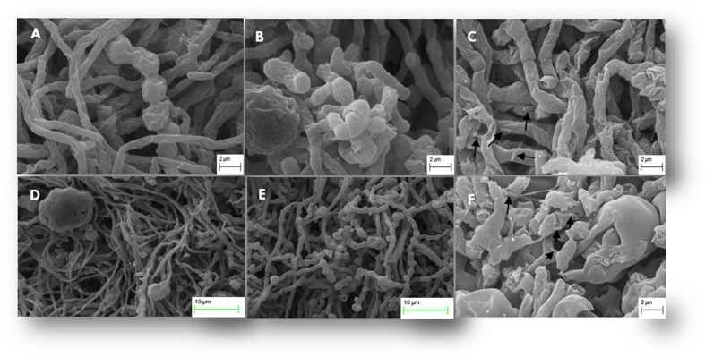
(A) and (D): Control fungal mycelia appear to have normal hyphae, septa and conidia. Hyphae showed uniform tubular shape in all parts. (B) and (E): micrographs show a tendency of hyphal growth promotion induced by WR5. The main improvements are healthy fungal hyphae and more conidiation. (C) and (F): Hyphal growth affected by M4 showing damaged fungal hyphae with surface adhered rod-shaped bacteria (arrow in Fig. 3C) and lack of conidiation. Data shown is the representative of at least three independent sets of experiments.