
Figure 3. Transmission electron micrographs of P. indica in isolation and in co-culture with A. chroococcum (WR5 and M4) grown at 28 ± 1 °C.
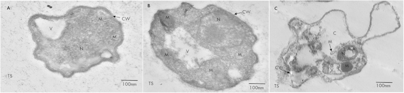
(A) Transverse section of control hypha showing cell wall (CW) mitochondria (M), vesicles (V) and nucleus (N). (B) Transverse section of hypha from co-culture with WR5 well organized hyphal cytoplasm, organelles and number of mitochondria. (C) Transverse section of hypha treated with M4 showing disorganization of hypha and cytoplasmic organelles and formation of membrane-bound vesicles. Data shown is the representative of at least three independent sets of experiments.