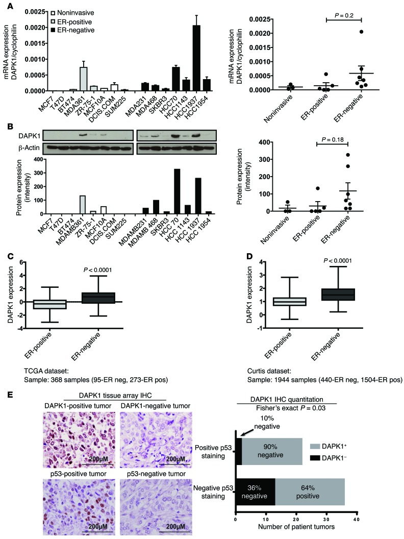
Figure 1. DAPK1 is differentially expressed in ER-negative and ER-positive breast cancers.
mRNA and protein expression of DAPK1 in ER-positive and ER-negative breast cell lines and tumors. Fifteen human breast cell lines and 2 data sets were used to analyze the expression of DAPK1 in breast tumors. (A) mRNA expression of DAPK1 in 15 human breast cell lines. Each cell line was done in triplicate, and 2-tailed Student’s t test was used for statistical analysis. (B) Protein expression of DAPK1 in 15 human breast cell lines. The quantification of DAPK1 expression levels in noninvasive, ER-positive, and ER-negative cell lines is shown in the right panel, with results reported as average ± SEM. Two-tailed Student’s t test was used for statistical analysis. (C) mRNA expression of DAPK1 in ER-positive compared with ER-negative breast tumors in TCGA data set (368 samples) (4). (D) mRNA expression of DAPK1 in ER-positive compared with ER-negative in the Curtis data set (1944 samples) (20). See also Supplemental Figure 1. DAPK1 expression data were grouped according to clinical receptor status annotations, and DAPK1 expression was compared using box-and-whisker plots and presented on a log2 scale. Two-tailed Student’s t test was used for statistical analysis. (E) Immunohistochemical detection of DAPK1 and p53 in human patient tissue microarray tumor cores and a comparison of the proportion of patients with or without p53 staining associated with DAPK1 levels (right panel). Original magnification, ×40. Comparison of immunohistochemical staining was evaluated using Fisher’s exact test.

