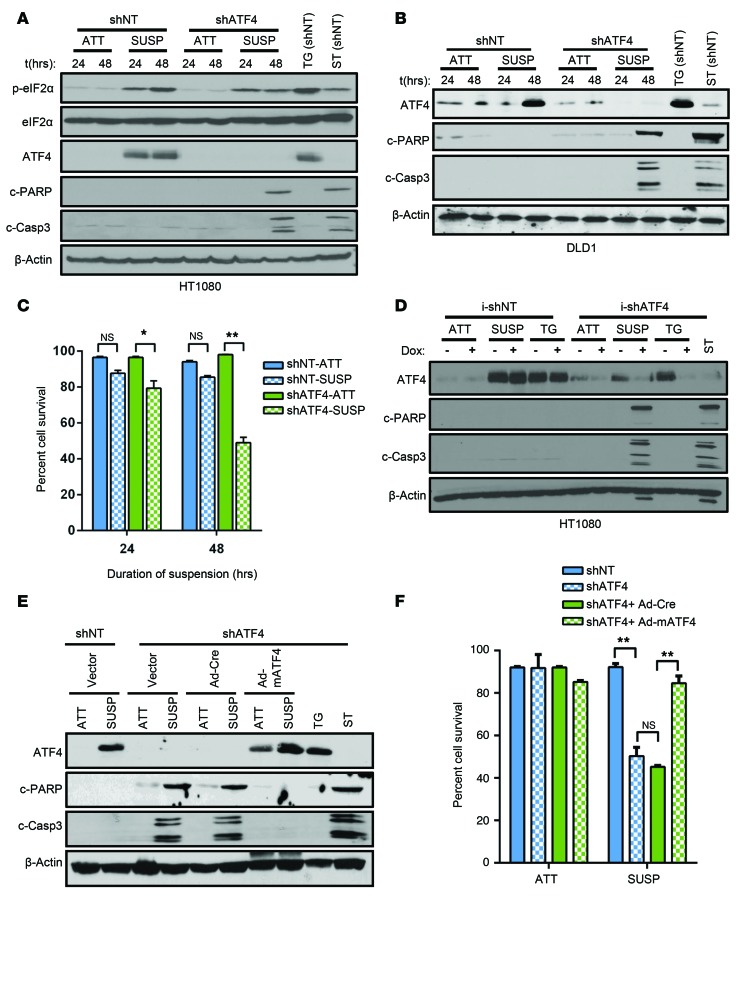
Figure 2. ATF4 confers resistance to anoikis.
shNT and shATF4.HT1080 (A) as well as shNT and shATF4.DLD1 cells (B) were grown in attached or in suspension conditions, and Western blot analysis for markers of the ISR and apoptosis was performed. (C) Mean percentage cell viability from 3 independent experiments was measured in shNT and shATF4.HT1080 cells grown in suspension (n = 3, mean ± SD). *P < 0.05; **P < 0.01, Student’s t test. (D) HT1080 cells stably expressing tetracycline i-shATF4 or i-shNT were grown in attached or suspension conditions for 48 hours or treated with 0.5 μM TG after pretreatment either with DMSO (–) or Dox (+) to alter ATF4 expression. Levels of c-PARP and cleaved caspase-3 were measured and compared with ST treatment. (E) Immunoblots of lysates from shATF4.HT1080 infected with empty vector, control (Ad-Cre), or mATF4 (Ad-mATF4) and grown in attached or suspension cultures for 48 hours. (F) Cell viability by Trypan blue exclusion assay. Percentage of cell survival is represented as mean ± SD from 3 independent experiments (n = 3, mean ± SD). *P < 0.05; **P < 0.01, Student’s t test. All immunoblots are representative of 2 independent experiments.

